AI System Uses Chest X-rays to Accurately Identify Collapsed Lungs
By MedImaging International staff writers
Posted on 04 Nov 2019
Researchers at the University of Waterloo (Waterloo, Canada) have developed a new assistive technology that can diagnose collapsed lungs, or pneumothorax, from chest X-rays with a higher degree of accuracy than radiologists. The system uses artificial intelligence (AI) software to search a huge database of X-ray images with known diagnoses for comparison to X-rays of new patients with unknown conditions. This approach enables the researchers to identify 75% of cases of collapsed lungs, as compared to medical specialists who diagnose less than 50% of cases using chest X-rays.Posted on 04 Nov 2019
The new AI software searches a database of more than 550,000 X-rays, including 30,000 cases of collapsed lungs, for those most similar to a new patient's X-rays. If the known condition in the majority of the similar X-rays is collapsed lung, then the AI recommends collapsed lung as the diagnosis in the new case.

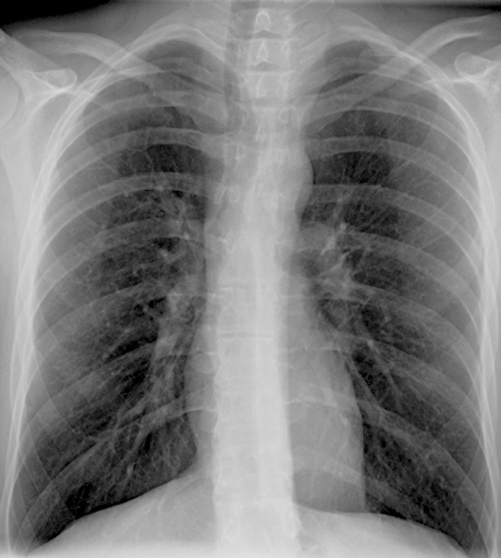
Image: An AI-enabled Coral Review would “scan through thousands of existing medical images (i.e., x-rays) for ones similar to a patient’s and recommend a diagnosis to the attending physician” (Photo courtesy of the Vector Institute).
The researchers are working with the University Health Network (UHN), a healthcare and medical research organization consisting of several Toronto-area hospitals, on a project backed by the Vector Institute, a not-for-profit corporation dedicated to advancing AI. The goal is to increase the accuracy of the technology to over 90% and integrate it next year into a software system, Coral Review, developed and used at UHN-affiliated hospitals. Coral, a quality assurance tool, allows doctors to provide second opinions by reviewing medical imaging diagnoses made by their peers. If the AI technology proves to be successful at UHN, then it would be offered to other hospitals, which are currently using Coral.
The system could offer radiologists a "computational second opinion" in Coral, or be used to prioritize the X-rays busy specialists look at first, reducing treatment delays that also put patients at risk. In addition to pneumothorax, the researchers also plan to apply the core AI search system to various other conditions, including pneumonia and chronic obstructive pulmonary disease (COPD), that are diagnosed using chest X-rays.
"Our results are very exciting," said Antonio Sze-To, a postdoctoral fellow at Waterloo. "The AI we use works almost like magic - and it will help radiologists save lives."
"There is no question systems like this will be in place in hospitals within the next two years," said Hamid Tizhoosh, a professor of systems design engineering and director of the Laboratory for Knowledge Inference in Medical Image Analysis (KIMIA Lab). "People are pushing for it and the technology is there."
Related Links:
University of Waterloo


