New AI Tool Could Radically Improve Diagnosis and Treatment of Breast Cancer
By MedImaging International staff writers
Posted on 27 Dec 2019
Researchers at the University of Auckland (Auckland, New Zealand) are combining machine learning and state-of-the-art imaging to develop an automated analysis technique that will radically improve the diagnosis and treatment of breast cancer. The researchers received USD 1.05 million in philanthropic funding to advance research in which they have developed biomechanical analysis techniques that automatically merges information from different medical images of the breast. This will provide clinicians with more information about any abnormality, as suspicious lesions and warning signs for cancer can appear differently in the various types of images.Posted on 27 Dec 2019
For instance, it can help to co-locate abnormalities such as micro-calcifications – tiny and difficult-to-detect features visible on X-ray mammograms that can indicate early stages of breast cancer – with regions of increased blood supply identified using MRI and which can also indicate tumor growth. Part of the challenge has been identifying the different biomechanical properties of the different types of breast tissues — to account for the individuality of each patient. The team was able to draw on 200 scans provided (with patient permission) by the Auckland District Health Board. The researchers have made enormous progress in developing methods for analysis and mathematical modelling of breast tissue.

Illustration
“This work is approaching real-time clinical application, which is very exciting in terms of realizing the benefits of advanced computational techniques in improving outcomes for patients,” said Dr. Anthony Doyle, an MRI expert at Auckland City Hospital, who has worked with the team on identifying key clinical challenges that need solving and providing feedback on their research.
Related Links:
University of Auckland

Gold Member
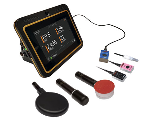
Solid State Kv/Dose Multi-Sensor
AGMS-DM+

New
Color Doppler Ultrasound System
KC20
Under Table Shield
3 Section Double Pivot Under Table Shield

New
X-Ray QA Meter
Piranha CT